Key Takeaways:
- Stanford researchers grew lab-developed human brain tissue inside cortex-depleted mice.
- Human cells replaced 92 percent of missing cortex and reached spinal cords.
- Scientists halted testing before human cells reached potential markers of consciousness.
Stanford University researchers have transplanted lab-grown human brain tissue in mice missing most of their cerebral cortex, creating a model to study human brain development and disease.
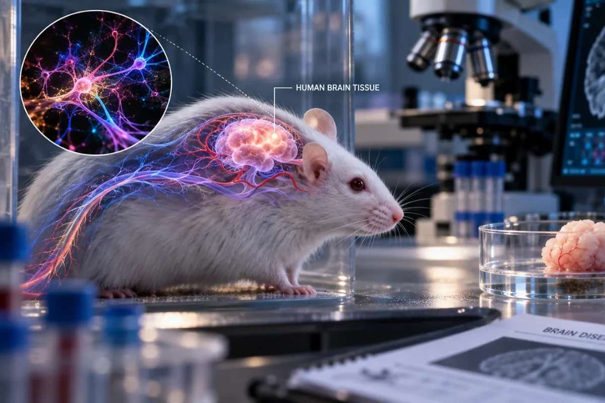
The study, published Sept. 16 in Nature, uses genetically engineered mice whose developing cortex was largely depleted before researchers implanted human cortical organoids. The human cells grew across most of the available space and formed connections with the mouse nervous system.
Human Cells Fill The Missing Cortex
The researchers created the mice by using a genetic strategy to remove most glutamatergic neurons from the developing neocortex and hippocampus before introducing human brain tissue in mice through transplanted cortical organoids.
More than 85% of implanted animals successfully incorporated the human tissue, according to the research. The human cells accounted for about 92% of the cells found in the animals’ cortex and developed several major types of cortical neurons.
The cells also sent long-distance projections through the mouse nervous system, with human-derived processes detected as far away as the spinal cord. Electrical recordings showed organized activity in the transplanted tissue, suggesting that the cells formed functioning neural circuits.
“These animal models offer a unique opportunity to study how disease-associated alterations in human brain circuitry manifest in an intact nervous system,” said Sergiu Pașca, a Stanford professor and senior author of the study.
The researchers also detected von Economo neurons, a rare cell type associated with parts of the human brain involved in social awareness and decision-making. The cells have been linked to frontotemporal dementia and have been difficult to produce in laboratory cultures.
Humanized Mice Show Mixed Results
The human tissue did not recreate a normal mouse cortex. Researchers found some local organization among cell types, but the transplanted cells did not form the distinct layers found in a typical cortex.
Behavioral tests produced mixed results. Mice with the human tissue performed better than cortex-free mice on a simple maze memory test but remained below normal mice. Their fine motor performance also fell between the two groups, while some associative memory tests showed little improvement.
The researchers also tested how the human tissue responded to oxygen deprivation. The altered mice developed walking and balance problems after exposure, while normal mice and cortex-depleted mice showed little effect.
“Finding out what accounts for this difference could yield clues about human neural susceptibility to oxygen deprivation,” Pașca said, adding that the findings could help researchers study conditions such as cerebral palsy.
Researchers See A Disease-Study Tool
The model of human brain tissue in mice could allow researchers to examine human brain cells inside a living nervous system rather than relying only on organoids grown in laboratory dishes.
The work also has ethical implications because it places substantial amounts of human neural tissue inside animals. Nita Farahany, a Duke University law and philosophy professor who served on an external ethics board for the project, said the researchers stopped the experiments before the human cells reached a developmental stage associated with a potential marker of consciousness.
The study of human brain tissue in mice does not create a complete human brain inside a mouse. The transplanted tissue lacks the normal layered organization of a cortex and remains part of a broader mouse nervous system. Researchers say further anatomical and behavioral studies are needed to determine how closely the model represents human brain function.