Introduction to New Imaging Technique
Traditional imaging methods such as light and electron microscopy have long been crucial in biological research. However, they each come with limitations—light microscopy struggles to resolve smaller features, and electron microscopy requires sample preparation that destroys living specimens. Atomic force microscopy (AFM) was developed to study the mechanical properties of materials at extremely high resolutions, but its slow imaging speed (taking several minutes per frame) was a barrier to observing living biological samples.
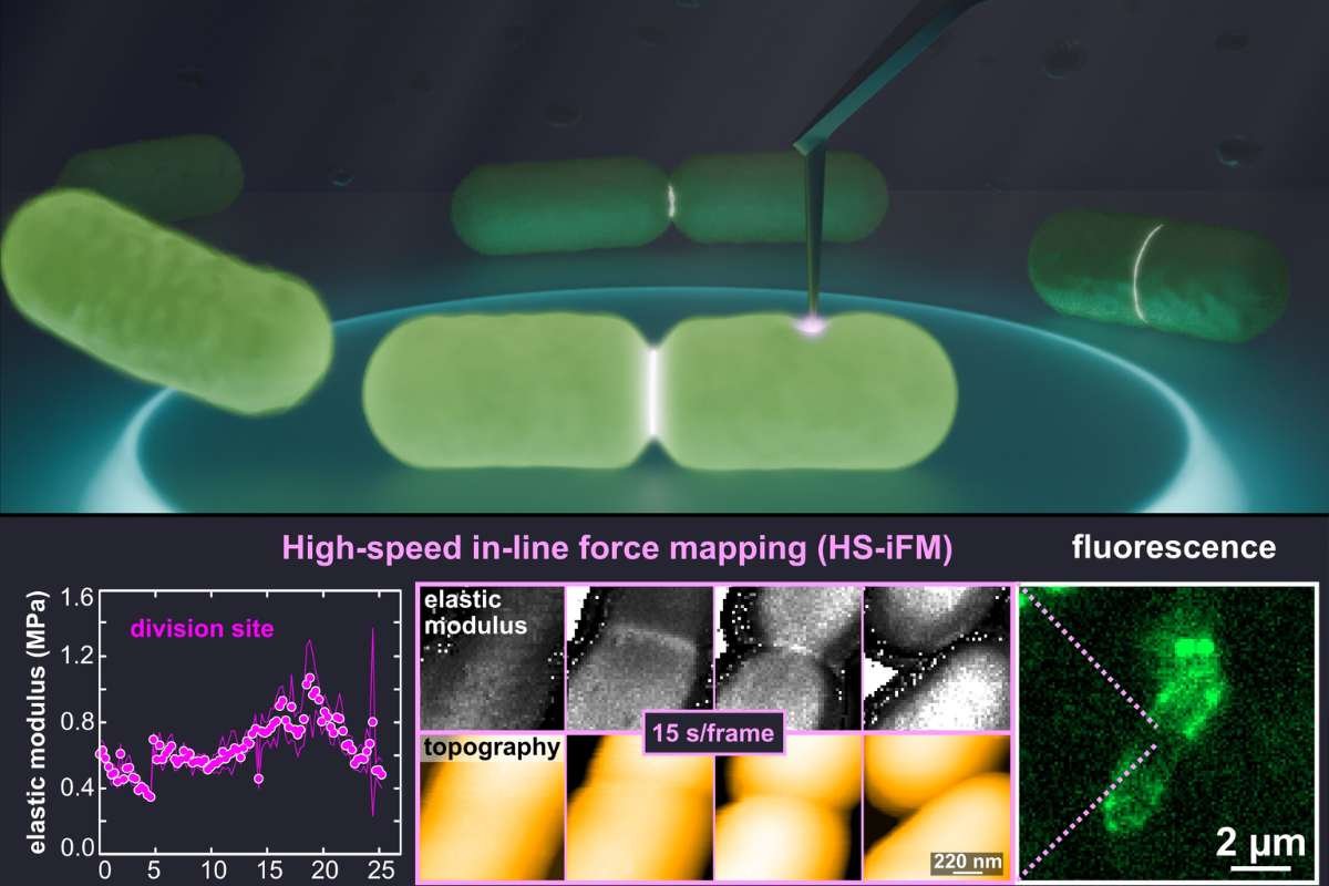
In response to this challenge, a team of researchers from the National Institutes of Natural Sciences (NINS) and Nagoya University have developed a novel technique called high-speed in-line force mapping (HS-iFM). This method combines the speed needed for live sample observation with the ability to capture detailed mechanical properties. In their latest study, the team used HS-iFM to examine the cell division dynamics of Escherichia coli, a model organism, in unprecedented detail.
Findings in E. coli Cell Division
The researchers’ observations revealed significant mechanical changes during the division of E. coli. They discovered that the division site of the bacteria became much stiffer than the surrounding cell areas. This increased stiffness likely resulted from localized tension within the membrane and thickening of the cell wall. According to Christian Ganser, one of the researchers, this stiffening suggests that internal stresses are required to deform the membrane and separate the two daughter cells. The team also observed visible bridges forming between the daughter cells during division. These membrane bridges stretched and eventually broke after an average of 242 seconds. This dynamic process, captured through HS-iFM, was also shared in a supplementary video accompanying the study.
Another key observation was the presence of a weak spot in the cell membrane, which ruptured during division, leading to cell depressurization and cell death. The rupture affected not only the dividing cell but also the daughter cells, implying that they were not fully separated internally at the time. This finding points to the potential of HS-iFM in determining the timing of various stages of bacterial division, which could be useful for understanding the process in other bacteria as well.
Applications and Future Prospects
The HS-iFM technique allows researchers to study both the topography and mechanical properties of biological membranes with high resolution. During the E. coli cell division, the team observed dynamic holes in the membrane, which would close, reform, and diffuse across the surface. These holes are thought to be related to the formation of outer membrane vesicles, which are more prevalent during cell division and the development of new cell walls between daughter cells. The size of these pores, around 34.7 nm, was much larger than previously reported for protein complexes, suggesting that the dynamics of bacterial cell division might be more complex than previously understood.
The researchers are excited about the potential of HS-iFM to study a wide range of biological samples beyond E. coli. Ganser noted that they plan to use the technique to explore the impact of external factors, such as antibiotics, on the nanomechanical properties of bacterial membranes. They also envision using HS-iFM to study the nanomechanical properties of polymers. Looking ahead, the team hopes to improve the technique’s speed and resolution to capture the mechanical properties of even smaller biological structures, such as individual proteins.