Key Takeaway:
- A 71-year-old woman developed two distinct primary pancreatic cancers within 12 months.
- Surgery followed by chemotherapy provided meaningful disease control despite later metastatic recurrence.
- The case highlights the need for expert review of surveillance scans and reassessment of new pancreatic tumours.
A 71-year-old woman developed two independent pancreatic cancers in 12 months in Moscow, with surgery and chemotherapy providing disease control and highlighting the need for careful surveillance.
First tumour responds to surgery
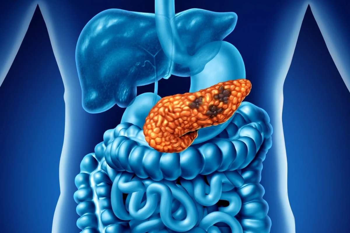
The case, reported by doctors at Sechenov University Clinical Hospital in Cancer Reports in June, involved a pancreatic neuroendocrine tumour followed by pancreatic ductal adenocarcinoma. Researchers classified the cancers as metachronous because they developed more than six months apart.
In 2022, imaging found a small mass in the head of the pancreas. The patient’s CEA and CA 19-9 tumour markers were normal. An endoscopic ultrasound-guided biopsy identified a well-differentiated, Grade 1 neuroendocrine tumour, with no evidence of distant spread.
Doctors performed a Whipple procedure — an operation refined by advances in multidisciplinary cancer care — removing the pancreatic head and nearby digestive structures. Pathology confirmed stage IA disease with clear surgical margins and no lymph node involvement, and the patient remained free of recurrence for 12 months.
The authors described the case as a rare example of “metachronous multiple primary pancreatic neoplasms,” emphasising that the two cancers had different histological characteristics.
Second cancer emerges in remaining pancreas
In May 2023, routine surveillance imaging detected a new mass in the tail of the remaining pancreas. Unlike the first tumour, the new lesion proved to be Grade 2 pancreatic ductal adenocarcinoma, with lymphovascular and perineural invasion and cancer in one of 10 lymph nodes.
The patient’s CA 19-9 level had risen to 1,700 units per millilitre, compared with a normal level during the first cancer. The findings showed why a new pancreatic lesion cannot automatically be assumed to represent recurrence of an earlier tumour.
Doctors performed a complete pancreatectomy with removal of the spleen and part of the left colon. The final diagnosis was stage IIB disease with clear margins. However, the surgery caused insulin-dependent diabetes, nutritional problems and severe weakness, preventing immediate postoperative chemotherapy.
Three months later, scans showed multiple liver lesions and other signs of metastatic disease. The patient began gemcitabine, a chemotherapy drug, after her functional status improved.
Chemotherapy controls disease before progression
The cancer remained stable during 11 cycles of gemcitabine over seven months. Imaging showed a 15% reduction in the combined size of the tracked liver lesions, although treatment caused severe thrombocytopenia, requiring a platelet transfusion and a missed dose.
The authors said the case demonstrated the value of a “histology-driven strategy” and multidisciplinary care in managing sequential pancreatic cancers. They also noted that progression visible on earlier scans was initially missed outside the tertiary centre, underscoring the importance of specialist review of surveillance imaging.
When the disease later progressed, the patient switched to irinotecan in March 2025. At the time of publication, she was tolerating the treatment with an ECOG performance status of zero to one, and further imaging was planned.
The researchers said the case supports closer reassessment of new tumours after pancreatic cancer treatment, including repeat pathological evaluation and, when appropriate, molecular testing, in line with National Cancer Institute guidance on post-treatment monitoring. They also called for greater use of technologies such as liquid biopsy and next-generation sequencing to help track tumour changes.
“Ensuring that surveillance imaging is reviewed by radiologists experienced in pancreatobiliary oncology is essential,” the authors wrote, warning that missed changes can delay treatment.