By cultivating “mini-placentas” in the lab, researchers have been able to learn more about the placenta’s development and interactions with the uterine lining. These discoveries may one day aid in the diagnosis and treatment of pre-eclampsia.
The research, which was published in Cell Stem Cell today, demonstrates that significant pregnancy abnormalities may be studied by conducting experiments on a developing human placenta as opposed to only seeing specimens.
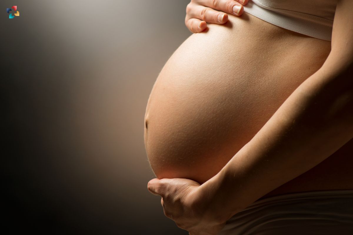
The placenta’s formation in the first few weeks of pregnancy is critical to a successful pregnancy. The placenta attaches itself in the endometrium, or the mucosal lining of the mother’s uterus, during this time.
The way that the cells of the placenta and the endometrium interact determines whether a pregnancy is successful. These interactions are particularly important for increasing the amount of maternal blood that reaches the placenta, which is crucial for the growth and development of the foetus.
Failure of these interactions to function correctly might result in issues like pre-eclampsia, which is a pregnancy-related illness characterised by elevated blood pressure. About six out of every 100 first pregnancies result in pre-eclampsia, which can be dangerous for the mother’s and the unborn child’s health.
Insight Into Pregnancy Problems Including Pre-Eclampsia
“Most of the major disorders of pregnancy – pre-eclampsia, still birth, growth restriction, for example – depend on failings in the way the placenta develops in the first few weeks. This is a process that is incredibly difficult to study – the period after implantation, when the placenta embeds itself into the endometrium, is often described as a ‘black box of human development.
Over the past few years, many scientists – including several at Cambridge – have developed embryo-like models to help us understand early pre-implantation development. But further development is impeded because we understand so little about the interactions between the placenta and the uterus.”
Professor Ashley Moffett, Department of Pathology, University of Cambridge
Using “mini-placentas,” a cellular model of the early stages of the placenta, Professor Moffett and colleagues at the Friedrich Miescher Institute in Switzerland and the Wellcome Sanger Institute in Cambridge have been able to shed light on early pregnancy and advance our knowledge of reproductive disorders. Tissue-grown from placenta cells, ‘trophoblast organoids’ resemble the early placenta so well that they have been found to produce a positive result on over-the-counter pregnancy tests.
Professor Moffett and colleagues have previously found genes that either protect against or raise the risk of illnesses like pre-eclampsia. These emphasised the critical function of immune cells that are only present in the uterus and are referred to as “uterine natural killer cells.” These cells gather in the womb’s lining near the location where the placenta implants. The interactions between the endometrial and placental cells are mediated by these cells.
Her team mimicked the conditions where the placenta installs itself in the trophoblast organoids by applying proteins released by the uterine natural killer cells. They discovered specific proteins that were essential for promoting the development of the organoids. By aiding in a successful implantation, these proteins will enable the placenta to enter the uterus and change the mother’s arteries.
Professor Moffett, who is also a Fellow at King’s College, Cambridge, stated, “This is the only instance that we are aware of where a normal cell invades and transforms an artery, and these cells are coming from another individual, the baby.”
The placenta and the unborn child are deprived of oxygen and nutrients if the arteries in the womb aren’t able to open up due to improper cell invasion. Because of this, you have issues later in pregnancy when the baby either dies or is very small because there is simply not enough blood to nourish it.”
Additionally, the researchers discovered a number of genes that control blood flow and aid in this implantation. According to Professor Moffett, these genes offer guidance for future studies aimed at improving our understanding of pre-eclampsia and related conditions.
Co-leading this effort with Dr. Margherita Turco of the Friedrich Miescher Institute in Switzerland, she continued, saying, “We still know relatively little about pre-eclampsia, despite the fact that it affects millions of women each globally. Pre-eclampsia typically manifests in women at the end of their pregnancy, but in order to truly comprehend, anticipate, and avoid it, we must examine the early stages of pregnancy.
Also Read: Early In Pregnancy, A “Groundbreaking” New Blood Test Can Identify Genetic Problems In Foetuses