The muscles of mastication play a vital role in the process of chewing or mastication, facilitating the breakdown of food particles and initiating the digestive process. Comprising a group of powerful muscles located around the jaw, these muscles are essential for proper oral function and overall health. In this comprehensive guide, we will delve into the anatomy, function, and common disorders associated with the muscles of mastication.
Anatomy of the Muscles of Mastication
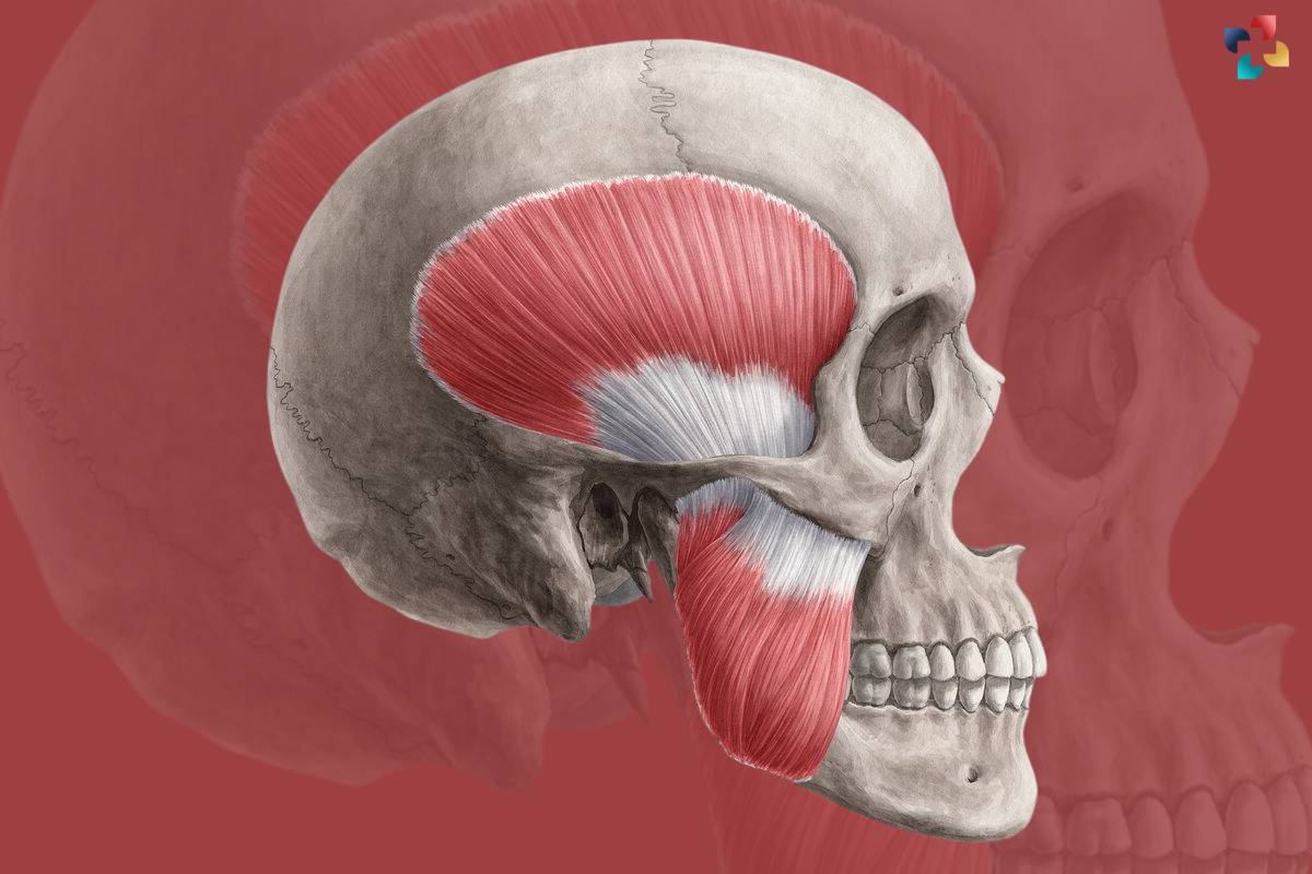
The muscles of mastication are primarily located in the head and neck region, centered around the temporomandibular joint (TMJ). The main muscles involved include the masseter, temporalis, medial pterygoid, and lateral pterygoid. These muscles work synergistically to coordinate the movements of the mandible (lower jaw) during chewing and other oral activities.
1. The Masseter Muscle
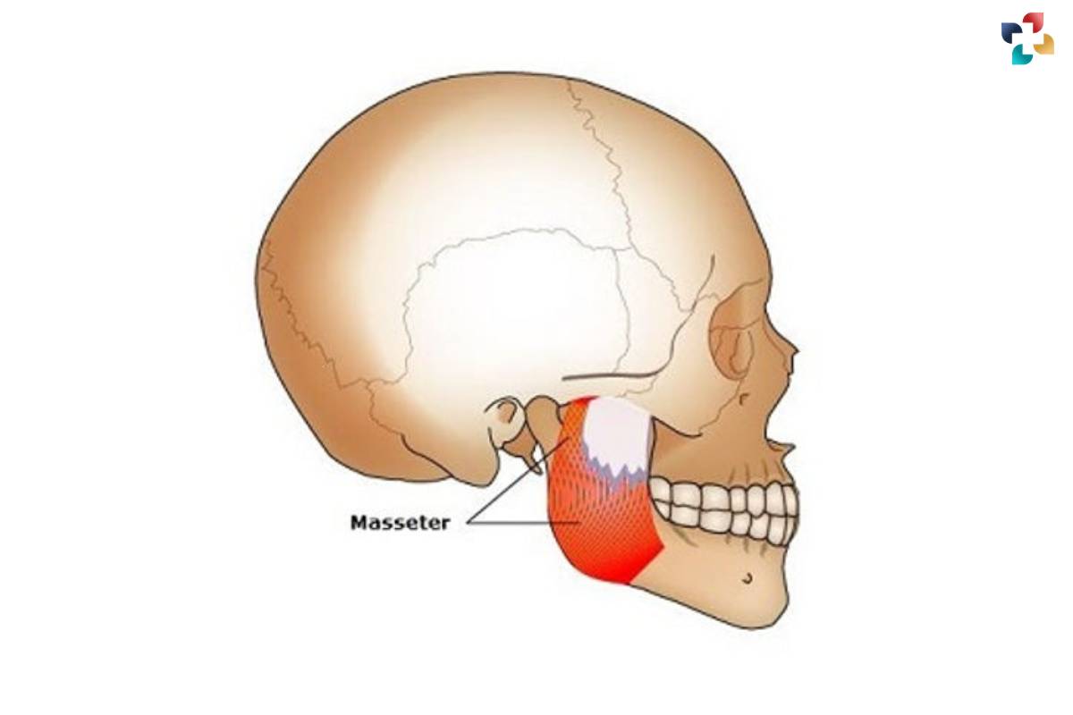
The masseter muscle is the largest and strongest muscle of mastication, originating from the zygomatic arch (cheekbone) and inserting into the mandibular ramus (jawbone). It plays a key role in elevating the mandible and closing the jaw during biting and chewing movements. The masseter muscle is divided into superficial and deep portions, with the superficial portion primarily responsible for jaw elevation and the deep portion aiding in jaw stabilization.
2. The Temporalis Muscle
The temporalis muscle is a broad, fan-shaped muscle located on the side of the skull above the ear. It originates from the temporal fossa (depression in the skull) and is inserted into the coronoid process of the mandible. The temporalis muscle functions to elevate and retract the mandible, assisting in the closing and posterior movement of the jaw during chewing. It also contributes to the side-to-side movements of the jaw, allowing for effective grinding of food particles.
3. The Medial Pterygoid Muscle
The medial pterygoid muscle is located deep within the mandibular region, originating from the medial surface of the lateral pterygoid plate of the sphenoid bone and inserting into the angle and ramus of the mandible. It works in conjunction with the masseter muscle to elevate the mandible and close the jaw. Additionally, the medial pterygoid muscle aids in protruding the mandible and moving it from side to side during chewing.
4. The Lateral Pterygoid Muscle:
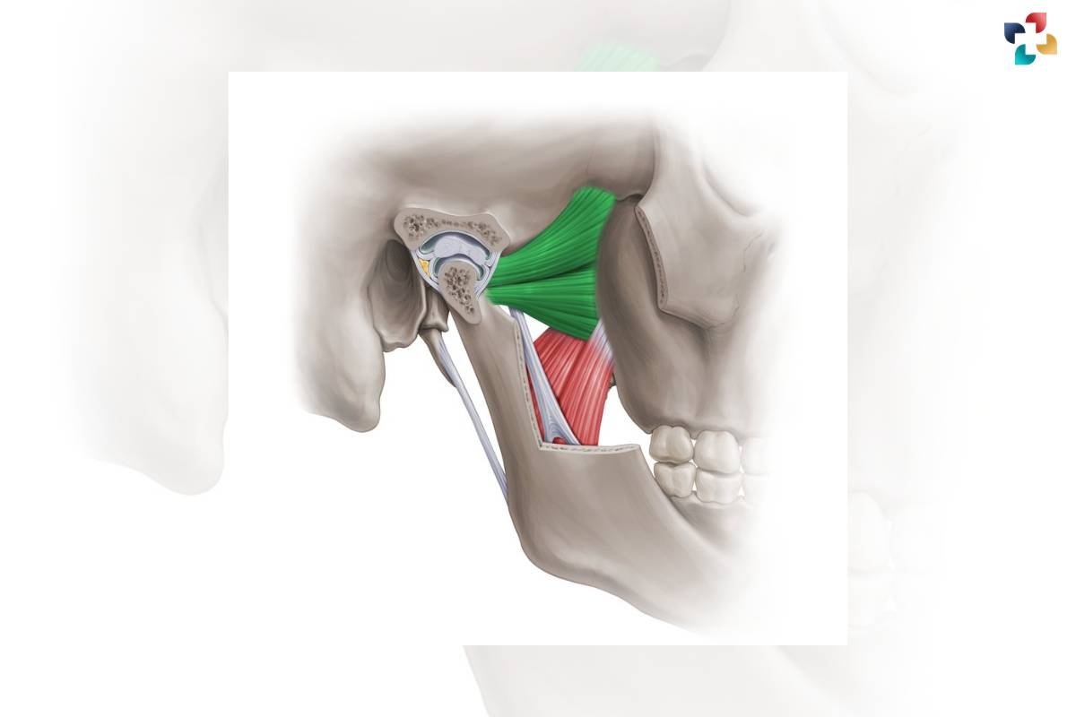
The lateral pterygoid muscle is situated deep within the temporomandibular joint (TMJ), originating from the greater wing of the sphenoid bone and inserted into the neck of the mandible. It plays a crucial role in opening the jaw by pulling the mandible forward and downward. Additionally, the lateral pterygoid muscle assists in protruding the mandible and moving it from side to side, contributing to the grinding and crushing of food particles during chewing.
Function of the Muscles of Mastication:
The primary function of the muscles of mastication is to facilitate the process of chewing or mastication. When food enters the mouth, these muscles contract and exert force on the mandible, causing it to move in various directions. The masseter, the strongest muscle of mastication, elevates and protracts the mandible, while the temporalis muscle assists in closing the jaw. The medial and lateral pterygoid muscles contribute to side-to-side movements and jaw stability during chewing.
Common Disorders of the Muscles of Mastication:
Despite their crucial role in oral function, the muscles of mastication are susceptible to various disorders and dysfunctions. Temporomandibular joint disorder (TMD) is one of the most common conditions affecting these muscles, characterized by pain, stiffness, and clicking sounds in the jaw joint. Bruxism, or teeth grinding, can also lead to muscle tension and soreness in it. Additionally, injuries, stress, poor posture, and malocclusion (misalignment of the teeth) can contribute to muscle strain and dysfunction.
Treatment Options for Muscles of Mastication Disorders:
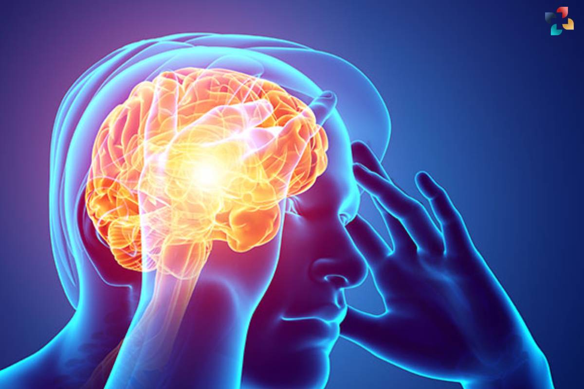
Treatment for disorders of the muscles of mastication typically focuses on relieving symptoms, reducing inflammation, and restoring normal function. Conservative approaches may include rest, applying heat or ice packs, physical therapy, and stress management techniques. In some cases, dental interventions such as occlusal splints or orthodontic treatment may be recommended to address underlying structural issues. For severe or persistent symptoms, medication, corticosteroid injections, or surgical procedures may be necessary to alleviate pain and improve jaw function.
Conclusion:
The muscles of mastication are integral to oral health and proper functioning of the jaw. Understanding their anatomy, function, and common disorders is essential for maintaining oral hygiene and addressing any issues that may arise. By adopting preventive measures and seeking timely treatment, individuals can ensure the optimal health and function of their muscles of mastication, promoting overall well-being and quality of life.
FAQs
1. What are the main muscles of mastication?
The main muscles of mastication include the masseter, temporalis, medial pterygoid, and lateral pterygoid muscles.
2. How do the muscles of mastication function during chewing?
The muscles of mastication work together to move the mandible (lower jaw) during chewing, helping to break down food particles into smaller pieces.
3. What causes temporomandibular joint disorder (TMD), and how does it affect the muscles of mastication?
TMD can be caused by factors such as jaw misalignment, teeth grinding (bruxism), stress, or trauma. It can lead to muscle tension, pain, and dysfunction in the muscles of mastication.
4. Can muscle tension in the muscles of mastication be relieved with home remedies?
Yes, applying heat or ice packs, practicing relaxation techniques, and avoiding excessive chewing or teeth grinding can help alleviate muscle tension in the muscles of mastication.
5. Are there exercises to strengthen the muscles of mastication?
Yes, exercises such as jaw opening and closing, side-to-side movements, and gentle massage of the jaw muscles can help strengthen and relax the muscles of mastication.

