Source – News-Medical.net
Why is what we now know about the development of the foetal immune system insufficient?
The roles of immune cells in somatic tissue development, homeostasis maintenance (especially in the gut and testis), and regeneration have all been well-documented in the past. Research has attempted to clarify the composition, subtypes, and functions of mesenchymal and epithelial cells; yet, there is still a lack of knowledge regarding the functions and mechanisms of lung immune cells.
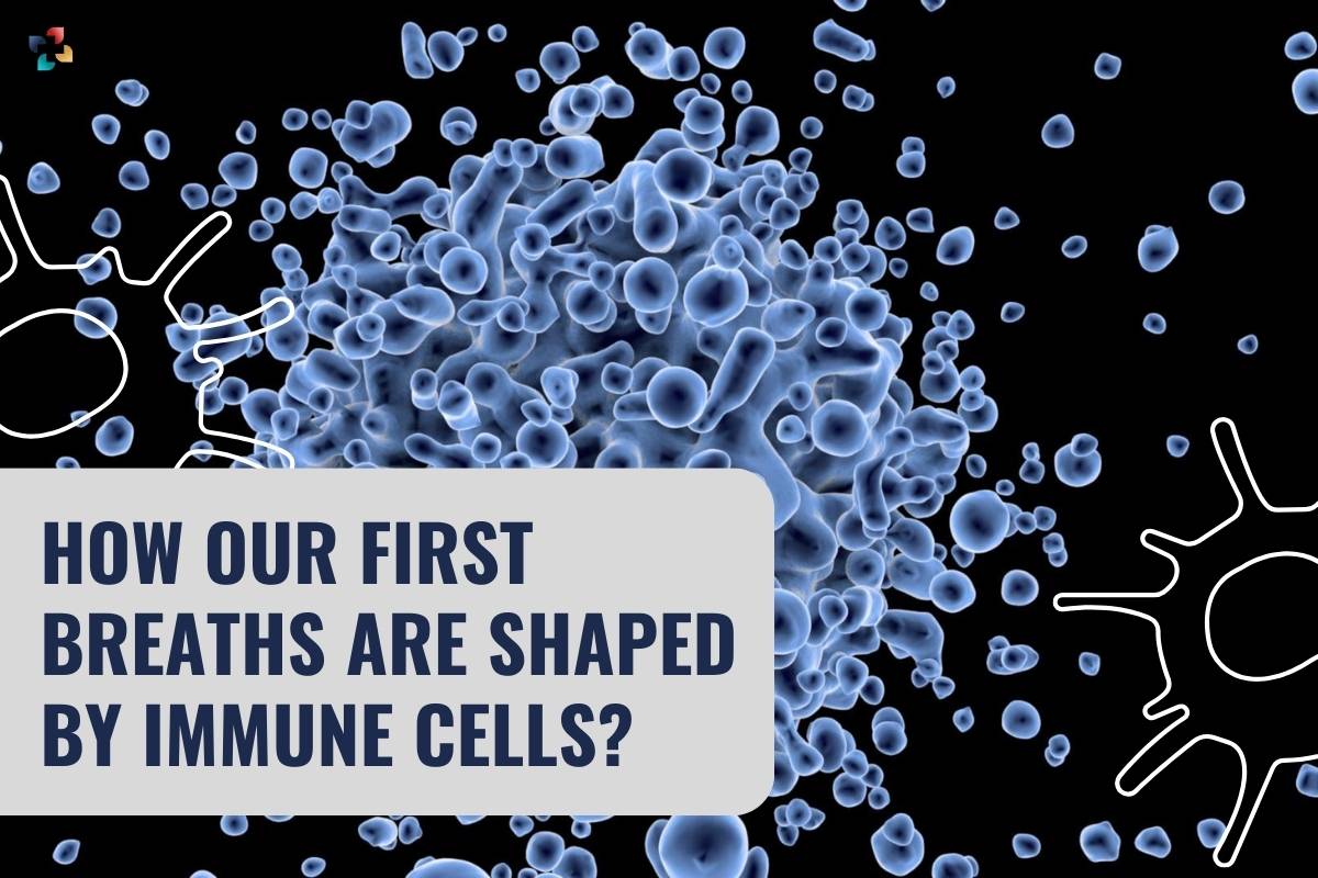
One of the most important cell types for an infant’s survival from the moment of birth is the immune system. It is remarkable that there is currently so little research on lung-associated mucosal immune cells’ defences against inhaled toxins and airborne infections. The intricate nature of cell differentiation during embryonic development and the historical dearth of methods capable of properly tracking these differentiations throughout pregnancy could be one reason for this gap in the literature.
Unanswered is a crucial question: may immune cells serve purposes other than defence? For example, by influencing the development of the tissues in which they live, or by modulating it in some other way? By providing answers to these and other related questions about human lung development at the cellular and molecular levels, millions or even hundreds of millions of patients will have an alternative to lung transplantation in the form of innovative clinical interventions intended to repair and regenerate lungs.
The morphology of human lung development has been described previously, and the process has been divided into five temporally overlapping stages. These include the following stages: the embryonic stage, which occurs between four and seven weeks after conception (pcw); the pseudoglandular stage, which occurs between five and seventeen pcw; the canalicular stage, which occurs between sixteen and twenty-six pcw; the saccular stage, which occurs between twenty-four and thirty-eight pcw; and the alveolar stage, which occurs between thirty-six pcw and twenty-one years of age.
Although the first three stages combined include the full evolution of epithelial stem cells into nearly functional lungs, the period between five and 22 pcw is the least studied period of lung development.
Concerning the study
The current investigation sought to assess the foetal immune system’s developmental trajectory and clarify how it might influence the development of the embryo’s lungs. Under the Human Developmental Biology Resource (HDBR) Joint MRC/Wellcome Trust grant, human foetal and embryonic samples were obtained.
With the donors’ signed consent, pregnancies ended between five and 22 postpartum weeks were used to harvest fresh lung tissue. The samples that were included were checked for genetic abnormalities and were shown to reflect ‘typical’ human embryonic growth using karyotypic analysis.
Validation of the types and numbers of immune cells during the first three stages of foetal lung development was achieved by immunohistochemistry (IHC) of lung tissues. The locations of the immune cells that were seen and their variations between five and 22 pcw were further evaluated with the help of IHC analysis.
Imaris software studies were carried out after three-dimensional (3D) quantification employing confocal microscopy to increase the precision and dependability of immune cell quantification. Throughout the course of the investigation, computed 3D images were compared to 2D photographs at every stage.
To validate IHC quantification estimates, determine the relative proportions of CD3+, CD4+, CD8+, and regulatory T cells (Tregs) as a percentage of CD45+ populations for the same developmental stage, and sort CD45+ cells as a precursor to single-cell ribonucleic acid (RNA) sequencing (scRNA-sq), lung tissue digestion was followed by flow cytometry and fluorescence-activated cell sorting (FACS).
Furthermore, the dual purposes of scRNA-sq were the molecular characterisation of immune cells across samples and the validation of flow cytometry data. To increase the resolution of the data, cellular indexing of transcriptomes and epitope sequencing (CITE-seq) was also used.
Functional characterisation studies involving cytokine treatments, dual Suppressor of Mothers Against Decapentaplegic (SMAD) transcription assays, macrophage, dendritic cell (DC) culture, and cytokine arrays were also performed on human embryonic lung organoids.
Using one-way Analysis of Variance (ANOVA), unpaired two-tailed t-tests, residual maximum likelihood analysis (REML), and Tukey’s post hoc multiple-comparison test, statistical analyses were performed on all collected data.
Human Immune System – How it works! (Animation)
Study results
The immune cell populations fluctuated dramatically during the foetal developing stages, as seen by the results of the scRNA-seq, IHC, and functional organoid assays. T- and B-lymphocytes eventually overtook progenitor and innate immune cells, such as myeloid, innate lymphoid (ILC), and natural killer (NK) cells, which dominated early developmental phases. Although CD45+ cells were almost universal in all lung-associated tissue areas and developmental stages, their relative abundance differed throughout time and space.
61,757 of the 77,559 transcriptome profiles that were discovered during the molecular characterization of immune cells are new to science. After these profiles were annotated and clustering analysis were performed, 59 clusters representing all known immune cell groups were identified. Unexpectedly large concentrations of ILCs and early lymphoid progenitor (ELP) were found through analyses of the progenitors of these categories.
When combined, these findings imply that immune cells develop in a biphasic manner during foetal development, peaking in quantity between eight and twenty post-conception weeks. Evaluations using quantitative polymerase chain reaction (qPCR) indicate that vascular maturation could be a contributing factor in the 20 pcw peak.
Through the use of IHC and single-molecule fluorescence in situ hybridization (smFISH) experiments, B-cell maturation in the lungs was first made clear. This contributed to the increasing amount of evidence suggesting, in contrast to earlier scientific theories, the bone marrow is not the only source of mature B-cells.
The expression of immunoglobulin (Ig) isotype in conjunction with clonal expansion experiments demonstrated that lung mesenchyme and epithelium secrete modulatory chemokines, such as CCL28, to maintain B-cell homeostasis during development.
The combined results of transcriptome and cytokine experiments demonstrated the intricate relationships between several cytokines released by immune cells, which were subsequently functionally confirmed to influence the development of epithelial cells. These findings support earlier theories by indicating that immune cells have a dual function during foetal development—that of defence and lung development.
In conclusion
The current work combines state-of-the-art transcriptome studies with immunohistochemistry (IHC) to clarify the structural and functional roles of immune cells during foetal and embryonic development. Analyses of foetal immune cells between five and thirty-three weeks post-fertility have shown that full B-cell maturation takes place in the embryonic lung, refuting the widely held notion that mature B-cell populations are exclusively found in the bone marrow.
It was discovered that widely scattered myeloid cells produced large amounts of interleukin-1 beta (IL-1β). The dual role of immune cells in lung epithelial growth and defence was highlighted by the discovery that IL-1β modulates and promotes epithelial stem cell differentiation.