In the ever-evolving world of medical and biological imaging, this is one of the emerging technologies. Offering high-resolution, label-free imaging, this advanced technique is making waves in various fields, from cell biology to clinical diagnostics.
In this blog, we will explore what this technology is, how it works, its applications, and the future it holds for science and medicine.
Definition
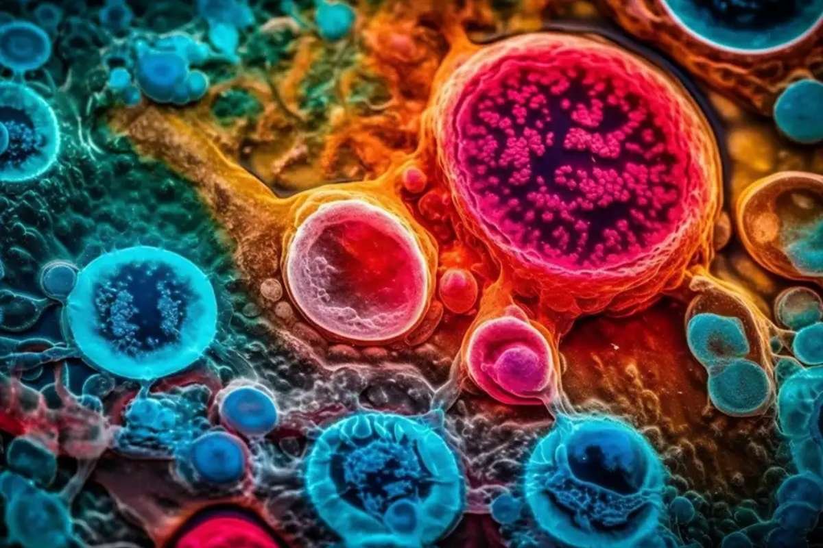
This is a cutting-edge imaging technique that combines principles from holography and tomography to produce three-dimensional (3D) reconstructions of biological samples at high resolution. Unlike traditional methods, holotomography does not require the use of fluorescent dyes or any other labels. This makes it a non-invasive, label-free imaging solution that can provide detailed information about living cells, tissues, and even entire organisms without altering their natural state.
The principle of this technology is based on the interference of light waves that pass through an object. By analyzing these light waves, it is possible to reconstruct a 3D image of the object. This technique allows researchers to observe the internal structure of cells and tissues with remarkable clarity, making it an invaluable tool for both basic research and clinical applications.
How this technology does Works?
It works by capturing interference patterns produced when light interacts with a sample. These patterns contain information about the refractive index of the sample, which can then be used to create a detailed 3D image. The technique is based on the principle of phase contrast imaging, which enables the visualization of transparent objects that do not naturally absorb or scatter light.
In a holotomographic system, a laser beam is directed through the sample, and the resulting light is captured by a detector. The data collected is processed by advanced algorithms to reconstruct the 3D image of the sample. The process is highly sophisticated, but it is also non-destructive, making it ideal for live-cell imaging and continuous monitoring.
Holotomography offers several advantages over traditional imaging techniques, such as optical microscopy or electron microscopy. It is faster, more versatile, and less invasive, making it particularly useful for studying living cells and tissues in real-time.
Applications
Holotomography is finding applications across a wide range of fields, particularly in biology, medicine, and materials science. Here are some of the most notable applications of this technology:
- Cell Biology: In cell biology, this technology is being used to observe living cells in 3D without the need for staining or labeling. Researchers can study cell morphology, growth, division, and interactions in real-time, providing valuable insights into cellular processes. This non-invasive approach is particularly beneficial for studying rare or delicate cell types that may be altered by conventional staining techniques.
- Cancer Research: This technology is proving to be a powerful tool in cancer research, allowing scientists to study the growth and behavior of cancer cells in 3D. By observing tumor cells in their native environment, researchers can gain a better understanding of cancer progression and response to treatments. This can lead to more effective therapies and personalized medicine approaches for cancer patients.
- Stem Cell Research: It is also being used in stem cell research to monitor stem cell differentiation and development. By providing a high-resolution, label-free view of stem cell behavior, researchers can gain deeper insights into how stem cells differentiate into various cell types. This has important implications for regenerative medicine and tissue engineering.
- Drug Discovery: The pharmaceutical industry is also benefiting from holotomography. By using this technology to analyze how cells respond to different drugs, researchers can better understand drug efficacy, toxicity, and mechanism of action. This can accelerate the drug discovery process and help identify potential treatments for various diseases.
- Clinical Diagnostics: This technology holds promise in clinical diagnostics, particularly for imaging tissues and organs in a non-invasive manner. By providing high-resolution images of tissues, this technology could assist in the early detection of diseases such as cancer, cardiovascular conditions, and neurological disorders.
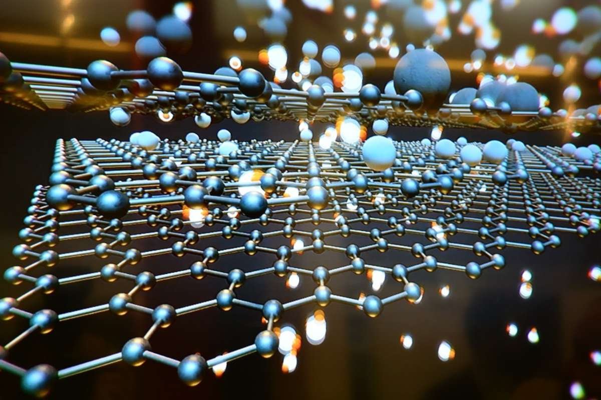
- Materials Science: Beyond biology and medicine, holotomography is also being applied in materials science. Researchers can use this technology to study the structure and properties of materials at the microscopic level. This is useful for a wide range of industries, including aerospace, automotive, and electronics.
Advantages
It offers numerous advantages over traditional imaging techniques, making it an increasingly attractive option for researchers and clinicians. Some of the key benefits of this technology include:
- Label-Free Imaging: One of the most significant advantages of holotomography is that it does not require any dyes or labels to visualize the sample. This eliminates the potential for artifacts that can occur when samples are chemically altered, ensuring that the observed structures are in their natural state.
- High Resolution: This technology provides high-resolution 3D images with exceptional clarity. This allows for the visualization of fine structures within cells and tissues, offering insights that would be difficult to obtain with conventional microscopy techniques.
- Non-Invasive: Since this technology does not require sample preparation or the use of chemical agents, it is non-invasive. This is particularly important for live-cell imaging, as it allows researchers to monitor dynamic cellular processes in real-time without damaging the cells.
- Real-Time Imaging: This technology allows for real-time imaging of living cells and tissues, making it possible to track biological processes as they unfold. This is particularly useful for observing cell behavior, such as division, migration, and response to stimuli.
- 3D Reconstruction: Unlike traditional microscopy, which provides only 2D imagesand generates 3D reconstructions of samples. This gives researchers a more comprehensive view of the sample’s structure, allowing for more accurate analysis and interpretation.
- High Throughput: Holotomography is a fast imaging technique, allowing for high-throughput analysis of large numbers of samples. This is particularly useful in drug screening and other applications that require the analysis of many samples in a short amount of time.
Challenges and Limitations
While holotomography is a powerful and promising technology, it does come with its own set of challenges and limitations. One of the primary challenges is the need for advanced computational algorithms to process the vast amounts of data generated during the imaging process. The reconstruction of high-resolution 3D images requires significant computational power, which can limit the scalability of holotomography in certain settings.
Additionally, holotomography may not be suitable for all types of samples. It is most effective for transparent or semi-transparent samples, and its performance can be limited when imaging highly dense or opaque materials. Further research is needed to optimize holotomography for a wider range of applications.
Future
As the technology continues to evolve, it is likely that holotomography will become an essential tool in both research and clinical settings. Advances in computational power, imaging hardware, and data analysis algorithms will improve the speed and resolution of holotomographic imaging, making it more accessible and practical for everyday use.
Conclusion
Holotomography is a great technology that is transforming the way we study biological systems. By providing high-resolution, label-free 3D imaging, it is enabling scientists and clinicians to gain deeper insights into cell biology, disease progression, and drug development. While challenges remain, the future of holotomography is bright, and its potential to revolutionize research and diagnostics is immense. As the technology advances, we can expect to see even more exciting applications in the years to come.