Table of Contents
Introduction to Epi-Fluorescence Microscopy
In the field of microscopy, epi-fluorescence microscopy is a potent instrument that provides unmatched clarity and precision in revealing the hidden world of fluorescent specimens. Wide-field fluorescence microscopy (WFM), which makes use of optical illumination and fluorescence, allows scientists to examine the complex dynamics and structures of biological molecules, cells, and tissues in extraordinary detail. We explore the mechanisms, benefits, and contributions to the scientific discovery of wide-field fluorescence microscopy (WFM) in this article as we set out to explore its amazing powers and uses.
Understanding Epi-Fluorescence Microscopy
Epi-fluorescence microscopy, also known as epifluorescence microscopy, is a specialized imaging technique that utilizes fluorescence to illuminate specimens of interest. Unlike traditional brightfield microscopy, where specimens are illuminated from below, epi-fluorescence microscopy employs a unique optical setup where both excitation and emission light pathways coincide, enhancing signal-to-noise ratio and contrast. This illumination configuration allows for selective excitation of fluorescent molecules within the specimen, while simultaneously blocking the excitation light from reaching the detector, resulting in highly sensitive and specific imaging.
Mechanisms of Epi-Fluorescence Microscopy
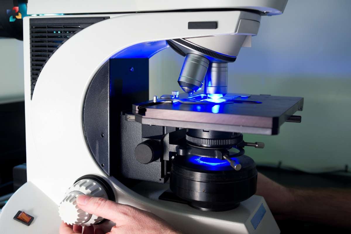
At the heart of epi-fluorescence microscopy lies the interaction between light and fluorescent molecules. When exposed to specific wavelengths of light, fluorescent molecules absorb photons and become excited to higher energy states. Upon returning to their ground state, these molecules emit photons of longer wavelengths, known as fluorescence emission. In wide-field fluorescence microscopy (WFM), a dichroic mirror is used to direct the excitation light towards the specimen while reflecting the emitted fluorescence towards the detector, enabling visualization of the fluorescent signal against a dark background.
Advantages of Wide-Field Fluorescence Microscopy (WFM)
Epi-fluorescence microscopy offers several distinct advantages over other imaging modalities, making it a preferred choice for various applications in life sciences and biomedical research. One of the key advantages is its ability to selectively image fluorescently labeled structures within complex biological specimens, such as cells or tissues, with high specificity and sensitivity. Additionally, epi-fluorescence microscopy enables real-time imaging of dynamic processes, allowing researchers to track molecular events and cellular behaviors with temporal precision. Furthermore, the non-invasive nature of fluorescence imaging minimizes phototoxicity and photobleaching effects, preserving specimen viability and enabling long-term observation.
Applications of Epi-Fluorescence Microscopy

The versatility of epi-fluorescence microscopy makes it indispensable for a wide range of biological and biomedical applications. In cell biology, wide-field fluorescence microscopy (WFM) is routinely used to visualize subcellular structures, study protein localization, and investigate molecular interactions within living cells. In neuroscience, epi-fluorescence microscopy facilitates imaging of neuronal morphology, synaptic activity, and neural circuit dynamics, providing valuable insights into brain function and dysfunction. Moreover, WFM finds applications in microbiology, immunology, developmental biology, and beyond, fueling discoveries in diverse fields of research.
1. Cell Biology:
Epi-fluorescence microscopy is utilized to study subcellular structures such as organelles (e.g., mitochondria, endoplasmic reticulum) and cellular compartments (e.g., nucleus, cytoplasm). Fluorescently labeled markers allow researchers to track the movement and dynamics of these structures in real time, providing insights into cellular processes such as mitosis, vesicle trafficking, and membrane dynamics.
2. Protein Localization:
By tagging proteins of interest with fluorescent labels, wide-field fluorescence microscopy (WFM) enables researchers to visualize their localization within cells. This approach is instrumental in determining the spatial distribution of proteins within cellular compartments and elucidating their roles in various biological processes, including signal transduction, gene expression, and cell signaling pathways.
3. Molecular Interactions:
Epi-fluorescence microscopy coupled with fluorescence resonance energy transfer (FRET) or fluorescence lifetime imaging microscopy (FLIM) allows researchers to investigate molecular interactions and protein-protein interactions in living cells. By monitoring changes in fluorescence intensity or fluorescence lifetime upon interaction, researchers can probe dynamic molecular events such as protein binding, conformational changes, and enzymatic activities.
4. Neuroscience:
In neuroscience, epi-fluorescence microscopy is widely used to study neuronal morphology, synaptic activity, and neural circuit dynamics. Fluorescent labeling techniques enable visualization of neuronal structures such as dendrites, axons, and synapses, facilitating studies on neuronal development, synaptic plasticity, and neuronal network function. Additionally, calcium-sensitive fluorescent dyes enable real-time imaging of neuronal activity and calcium dynamics in live brain tissue.
5. Microbiology:
Epi-fluorescence microscopy plays a crucial role in microbiology by enabling the visualization and study of microbial cells, pathogens, and microbial communities. Fluorescent labeling techniques allow researchers to track the behavior and interactions of bacteria, viruses, and fungi in complex environments. Epi-fluorescence microscopy is also employed in studies on microbial biofilms, antibiotic resistance, and microbial ecology.
6. Immunology:

In immunology, wide-field fluorescence microscopy (WFM) is used to study immune cells, immune responses, and host-pathogen interactions. Fluorescently labeled antibodies enable visualization of specific immune cell populations (e.g., T cells, B cells, macrophages) and their activation states in tissues and organs. This approach facilitates investigations into immune cell dynamics, immune cell trafficking, and immune-mediated diseases such as inflammation and autoimmunity.
7. Developmental Biology:
Epi-fluorescence microscopy is instrumental in studying embryonic development, organogenesis, and tissue morphogenesis. Fluorescent labeling techniques allow researchers to track cell lineage, cell migration, and tissue patterning during development. This approach provides insights into the molecular mechanisms underlying developmental processes and contributes to our understanding of birth defects, regeneration, and tissue repair.
These examples illustrate the diverse applications of epi-fluorescence microscopy in advancing our understanding of biological processes and addressing key questions in various fields of research.
Challenges and Future Directions
Despite its many advantages, epi-fluorescence microscopy is not without challenges. One limitation is the potential for autofluorescence, where endogenous molecules within the specimen emit fluorescence signals, complicating image interpretation. Additionally, the reliance on fluorescent labels necessitates careful selection of fluorophores and optimization of imaging conditions to minimize background noise and maximize signal-to-noise ratio. Looking ahead, advancements in fluorescent probe design, imaging instrumentation, and computational analysis tools are poised to further enhance the capabilities of wide-field fluorescence microscopy (WFM), opening new frontiers in biological imaging and discovery.

Comprehensive Cellular Mapping of a Mammalian Brain: A Neuroscience Milestone
The development of precise medicines for brain illnesses and a deeper understanding of the human brain are made possible by a ground-breaking cell atlas that details over 32 million cells in the mouse brain.
Conclusion
In summary, epi-fluorescence microscopy is a key tool in contemporary biological and biomedical research because it enables researchers to examine the hidden domains of fluorescence with a depth and clarity never before possible. Wide-field fluorescence microscopy (WFM) is transforming our knowledge of the living world by helping to grasp everything from the intricacies of disease pathophysiology to the secrets of cellular dynamics. Researchers light the way for ground-breaking discoveries and game-changing breakthroughs by utilizing light and fluorescence, paving the way for a healthier and brighter future.