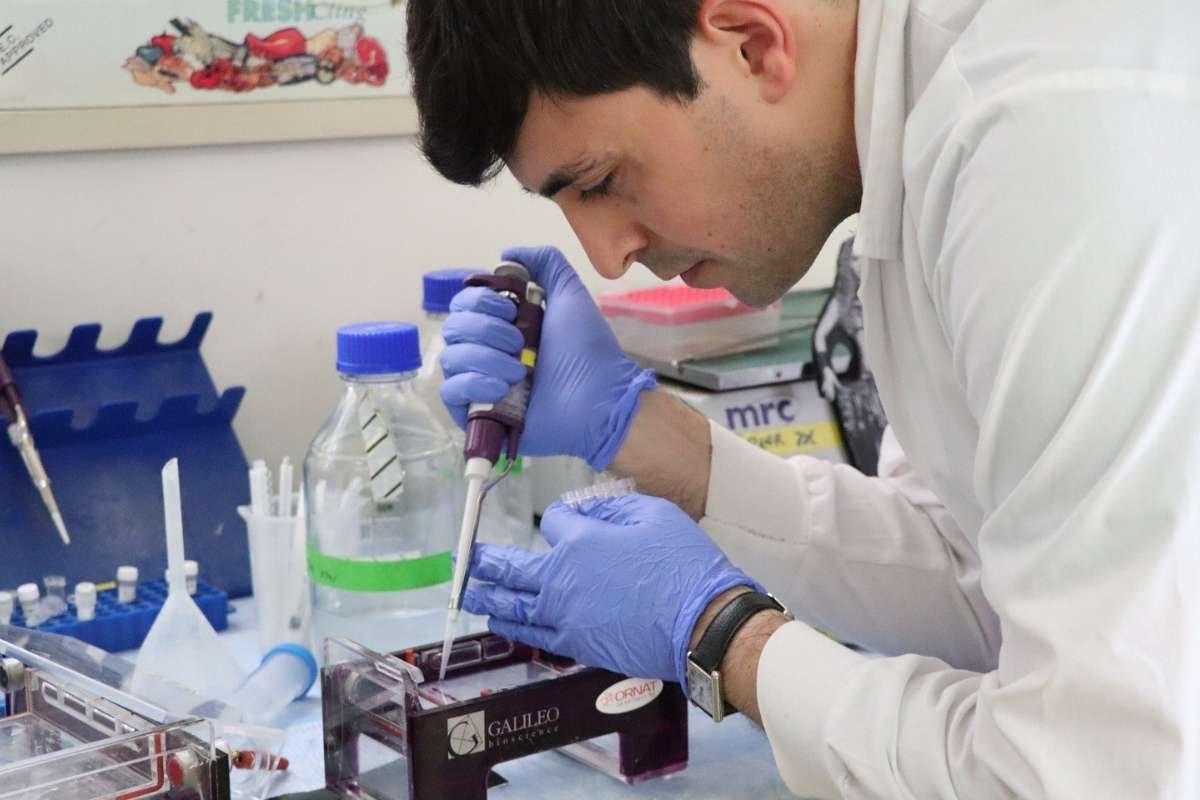
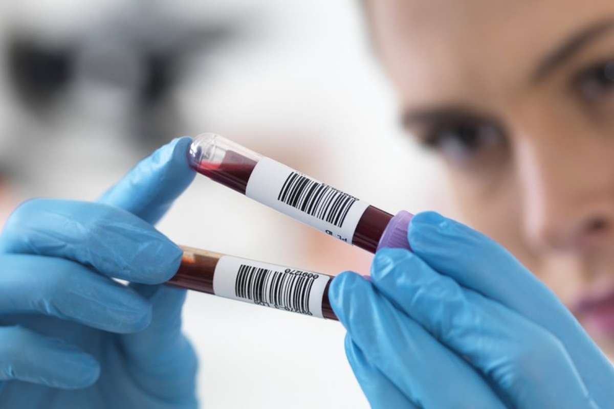
Being a ground-breaking field, digital pathology combines medical science, computer technology, and imaging. It has revolutionized the way pathologists and researchers analyze samples of tissue. Whole Slide Imaging (WSI) and Tissue Image Analysis are two pivotal aspects of digital pathology that have transformed the traditional practice of pathology. In this article, you will explore the world of digital pathology in-depth and read about the key components of Whole Slide Imaging along with Tissue Image Analysis. This article will highlight the significance of digital pathology in modern healthcare and research.
Understanding Digital Pathology
Digital Pathology is the confluence of pathology that combines studying various diseases and conditions with advanced digital imaging technology. It entails the acquisition, management, and interpretation of high-resolution digital images of pathological specimens, such as tissue samples, blood smears, and other cytological preparations. To get such digital images you can use specialized scanners to analyze, store, and share electronically. Digital pathology has wide-ranging applications, from clinical diagnosis and medical research to education and remote consultations.
Whole Slide Imaging (WSI)
Also referred to as virtual microscopy, Whole Slide Imaging (WSI) is a fundamental part of digital pathology. It involves the high-resolution scanning of entire glass slides containing pathological specimens, resulting in digital replicas of the entire slide. Such kind of digital representations can capture a similar level of detail and clarity as traditional optical microscopy has.
Key Aspects of Whole Slide Imaging:
- High-Quality Scanning: To ensure high-quality digital replicas and capture images of entire glass slides at different magnifications, WSI scanners use precision optics and sophisticated imaging sensors.
- Storage and Accessibility: Digital slides are stored electronically, eliminating the need for physical storage space and enabling easy access and retrieval of cases for reference and research.
- Remote Viewing: It is one of the important key aspects of WSI as pathologists and researchers review digital slides from anywhere with the help of an internet connection. It makes remote consultations, collaboration, etc,
- Annotation and Marking: WSI software allows pathologists to annotate, mark, and highlight specific areas of interest within the digital slide, aiding in research and educational purposes.
- Image Analysis: Automated image analysis of digital slides can help psychologists identify and quantify specific features for example cell counts or tissue patterns.
- Archiving and Backup: Digital slides can be archived and backed up, reducing the risk of slide damage or loss due to environmental factors.
Significance of Whole Slide Imaging:
- Efficiency and Accuracy: WSI is capable of streamlining the review and analysis of the pathological specimens leading to speedy diagnoses and potentially more accurate results.
- Education and Training: Digital slides provide a valuable resource for medical education and training, allowing students and trainees to access a vast collection of cases.
- Remote Consultations: Because of Whole Slide Imaging, pathologists can collaborate and seek the opinions of experts from specialists around the world instead of physical slide shipping.
- Research Advancements: WSI facilitates large-scale research projects by enabling the analysis of extensive slide archives and the application of image analysis algorithms for discoveries in pathology.
Tissue Image Analysis
Tissue Image Analysis involves the process of abstracting useful information from digital images of pathological specimens. It comprises applying computer algorithms and machine learning techniques to identify relevant structures, analyze tissue images, and support other objective measurements. This approach is particularly valuable in research, diagnostics, and treatment planning.
Key Aspects of Tissue Image Analysis:
- Object Recognition: Tissue Image Analysis software can identify and outline particular regions or objects of interest within an image, such as cells, nuclei, or tissue structures.
- Feature Measurement: The software of Tissue Image Analysis measures different features that include size, shape, texture, and staining intensity of objects within the image.
- Pattern Recognition: Pattern recognition is one of the key aspects of Tissue Image Analysis. Its algorithm can identify numerous complex patterns inside tissues and enable the recognition of abnormal tissue structures or cellular arrangements.
- Quantitative Data: Tissue image analysis generates quantitative data, such as cell counts, mitotic rates, and the extent of tissue damage, which can assist in diagnosis and research.
- Reproducibility: By automating the analysis process, Tissue Image Analysis ensures consistency and reproducibility in quantifying pathological features.
- Scalability: Tissue Image Analysis can be applied to a large number of images, making it suitable for high-throughput research and clinical applications.
Significance of Tissue Image Analysis:
- Objective Diagnosis: Tissue image analysis provides quantitative data and identifies subtle pathological changes that help pathologists make more objective and consistent diagnoses.
- Predictive Markers: Image analysis can uncover predictive markers for disease progression, treatment response, and patient outcomes, aiding in personalized medicine.
- Drug Development: Researchers all around the world use Tissue Image Analysis to assess the impacts of potential therapeutic agents on pathological tissue in drug development studies.
- Image-Based Biomarkers: It enables the discovery and validation of image-based biomarkers that can have clinical significance in disease diagnosis and prognosis.
- Research Efficiency: Tissue image analysis speeds up the whole research by automating the analysis of large datasets and leads to more rapid discoveries in pathology in the future.
Applications of Digital Pathology, WSI, and Tissue Image Analysis:
The integration of digital pathology, Whole Slide Imaging, and Tissue Image Analysis has brought about transformative changes in several domains:
1. Clinical Diagnostics
In clinical settings, digital pathology has wide applications to increase the accuracy and efficiency of diagnosing diseases, including cancers and diabetes. For remote consultations and automated image analysis, pathologists can access digital slides and assist in identifying anomalies, grading tumors, and predicting patient outcomes.
2. Medical Education
Digital pathology serves as a valuable resource for medical education and training. Medical students, residents, and professionals can access a vast repository of digital slides for learning and skill development.
3. Research and Drug Development
Tissue Image Analysis is at the center of pharmaceutical research. It aids in evaluating the effects of potential drugs and potential tissue and discovering new biomarkers to get insights into the mechanism of disease. Researchers can assess large image datasets within no time and enable high-throughput studies.
4. Telepathology
Telepathology, facilitated by Whole Slide Imaging, allows pathologists to consult with peers or experts across geographical distances. This technology is especially critical in areas with limited access to pathology expertise.
5. Bio Banking
In the field of bio banking, digital pathology plays a crucial role as with the help of this you can preserve the tissue samples for research activities in the future. Scanning these samples as digital slides ensures that they remain accessible for analysis and study.
6. Precision Medicine
Tissue image analysis contributes to the upgradation of precision medicine while identifying image-based biomarkers. To tailor treatments of individual patients these biomarkers can be used that can lead to more effective therapies.
Challenges and Future Prospects
In the world of healthcare, digital pathology, WSI and Tissue Image Analysis have contributed to making significant strides, however, a few concerns still exist in the field. It includes challenges regarding data security, the requirement of standardized practices, and the integration of digital pathology systems into healthcare infrastructures. We can expect these things from digital pathology as technology is thriving to advance in the industry:
- AI Integration: The greater amalgamation of artificial intelligence and machine learning algorithms in the area of Tissue Image Analysis leads to more efficient and accurate pathology assessments.
- Enhanced Collaboration: Improved tools for remote collaboration, enabling pathologists to work together seamlessly from different locations.
- Regulatory Developments: Upgradation in the regularity guidelines can ensure that it has a safe and effective implementation of digital pathology in healthcare.
- Global Accessibility: Wider adoption of digital pathology solutions, making them accessible to healthcare facilities in diverse settings.
- Innovative Applications: With the help of digital pathology, WSI, and Tissue Image Analysis, you can explore the innovative applications of digital pathology, for example, 3D reconstruction of tissue structures for deeper insights into pathology.
Conclusion
Having a main focus on Whole Slide Imaging and Tissue Image Analysis, digital pathology has been inaugurated in a new era of the field of pathology in which medical research is a crucial part. It has improved the efficiency and accuracy of disease diagnosis, provided an invaluable resource for medical education and research, and contributed to the development of precision medicine. As technology continues to advance and challenges are addressed, digital pathology is poised to play an increasingly central role in the future of healthcare and pathology, leading to better patient outcomes and innovative research discoveries.
Also Read: Is Telehealth the Present and Future of Healthcare?


