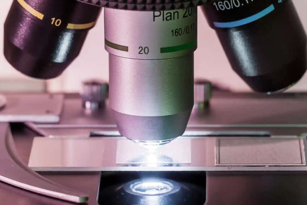
Unveiling Molecular Dynamics with Raman Microscopy
Understanding molecular and cellular behavior is pivotal for advancements in medicine, and researchers continue to strive for imaging techniques that reveal intricate biological processes. In a groundbreaking study published in Science Advances, scientists from Osaka University have introduced a significant improvement in Raman microscopy, a tool known for its ability to provide chemical insights into biological samples. This innovation holds the potential to revolutionize how researchers study molecules like proteins involved in critical bodily processes.
Traditionally, Raman microscopy faces limitations due to the weak light signals emitted by biological samples, which are often overpowered by background noise. This drawback has hindered its ability to deliver clear, high-resolution images. However, the team from Osaka University has tackled this issue with a novel method that enhances signal clarity, paving the way for a more precise understanding of cellular and molecular behavior.
A Leap Forward: The Cryofixed Microscope
Central to this advancement is a specially designed microscope that maintains the temperature of previously frozen biological samples during imaging. This cryofixed approach allows the production of images up to eight times brighter than those achieved using conventional Raman microscopy methods. Unlike other techniques, this method does not rely on chemical stains or fixation, ensuring an authentic representation of cellular processes and molecular behavior.
Additionally, the freezing process has been shown to preserve the physicochemical states of proteins, a feat chemical fixation methods struggle to achieve. This preservation not only enhances the reliability of the imaging process but also makes Raman microscopy a more versatile tool in biological research.
“Raman microscopy adds a complementary option to the imaging toolbox,” noted senior researcher Katsumasa Fujita. “Its ability to provide cell images alongside detailed information about molecular distribution and chemical states is invaluable for deepening our understanding of biological systems.”
Implications for Medicine and Pharmaceutical Sciences
The improved Raman microscopy technique offers far-reaching implications for the biological sciences, particularly in medicine and pharmaceutical research. By combining this method with other microscopy technologies, scientists can achieve a more comprehensive analysis of biological samples. The detailed chemical insights provided by this approach are expected to drive progress in fields such as drug development, disease diagnostics, and cellular biology.
This breakthrough represents a significant step forward in non-invasive imaging technologies, enabling researchers to uncover the complex mechanisms governing life at the molecular level. With the integration of this advanced Raman microscopy method, the future of biological imaging looks brighter than ever, promising enhanced understanding and innovation across a wide array of scientific disciplines.