Source-Verywell Health
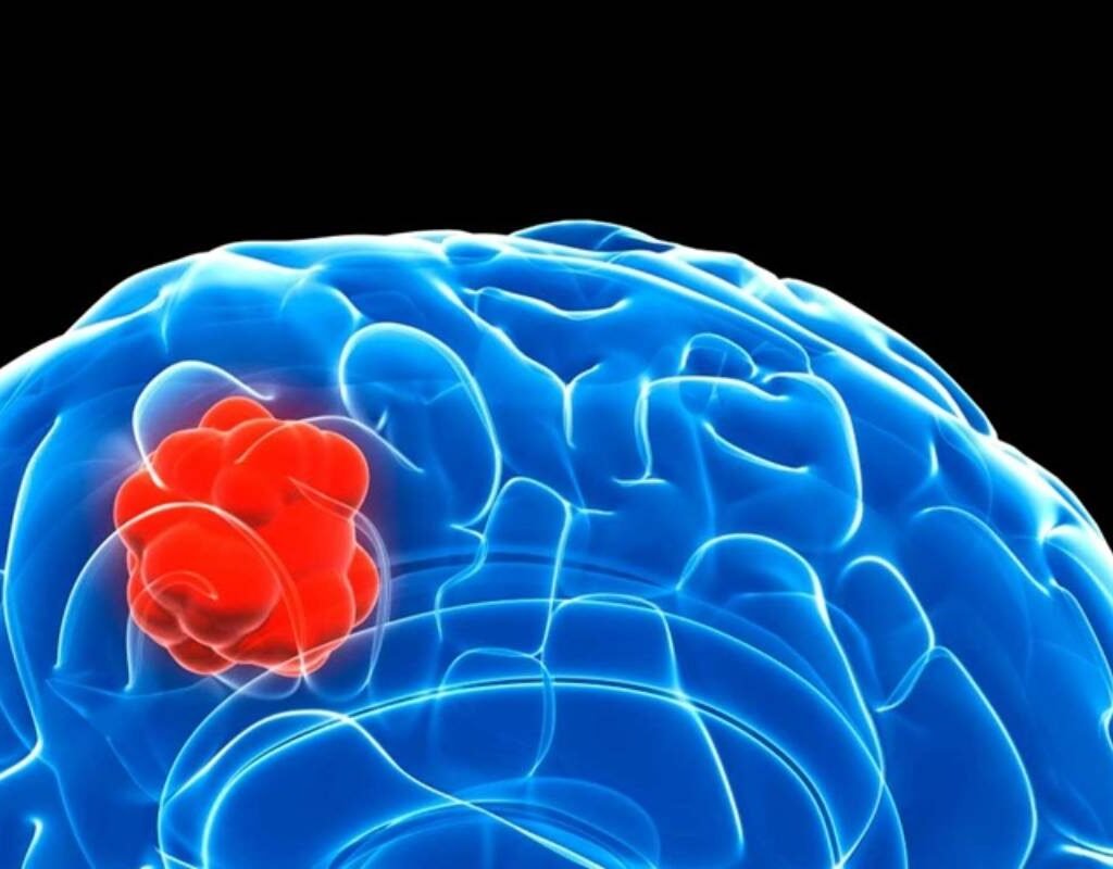
One of the most prevalent primary brain tumors is meningioma, which develops from the meninges, the membranes that surround the brain and spinal cord. Despite their generally benign nature, meningiomas can, depending on their size, location, and development pace, result in serious neurological symptoms and problems. We explore the clinical presentation, diagnostic techniques, treatment options, and the most recent developments in research and management tactics as we delve into the intricacies of meningioma in this thorough guide.
Understanding Meningioma: Origins and Characteristics:
Meningioma arises from the arachnoid cells of the meninges, predominantly occurring in the dura mater, the outermost layer of the meninges. These tumors are typically slow-growing and may remain asymptomatic for years, posing challenges in early detection and diagnosis. Meningiomas are classified based on their histological features, with the World Health Organization (WHO) grading system categorizing them into grades I, II, and III, depending on their likelihood of recurrence and malignant potential.
In addition to their classification based on histological features, meningiomas can also exhibit a diverse range of morphological characteristics, including their size, shape, and growth patterns. While most are solitary tumors, occasionally, they may present as multiple lesions, raising concerns about potential metastasis or underlying genetic predisposition syndromes. Furthermore, advancements in molecular profiling have revealed distinct molecular subtypes, providing insights into their underlying genetic alterations and biological behavior. Understanding the origins and characteristics at both histological and molecular levels is essential for accurate diagnosis, prognostication, and selection of optimal treatment strategies.
Clinical Presentation and Symptoms of Meningioma:

The clinical presentation of meningioma varies depending on factors such as tumor size, location, and proximity to critical brain structures. Common symptoms may include headaches, seizures, focal neurological deficits, cognitive impairment, and changes in vision or hearing. The onset and progression of symptoms may be gradual, leading to delayed diagnosis and treatment initiation. In some cases, meningiomas may compress adjacent brain tissue or blood vessels, causing intracranial pressure and neurological dysfunction.
Diagnostic Modalities for Meningioma Detection:
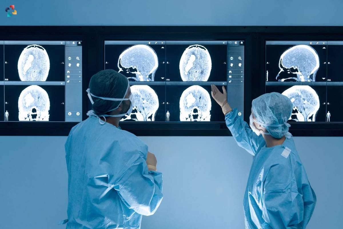
The diagnosis typically involves a combination of imaging studies and histopathological evaluation. Magnetic resonance imaging (MRI) is the imaging modality of choice for visualizing, providing detailed anatomical information, and delineating tumor characteristics such as size, location, and vascularity. Computed tomography (CT) scans may also be used to assess bony involvement and calcifications associated with it.
Histopathological examination of tissue samples obtained through biopsy or surgical resection confirms the diagnosis and provides valuable insights into tumor grade and subtype. In addition to MRI and CT scans, advanced imaging techniques such as magnetic resonance spectroscopy (MRS) and perfusion-weighted imaging (PWI) can provide valuable functional information about, such as metabolic activity and blood flow characteristics. These techniques aid in differentiating from other intracranial lesions and assessing tumor aggressiveness. Moreover, positron emission tomography (PET) scans with radiolabeled tracers targeting specific biological processes, such as glucose metabolism or cell proliferation, may offer complementary information for tumor characterization and treatment planning. Multimodal imaging approaches, combined with clinical and pathological data, enhance the accuracy of diagnosis and facilitate personalized management strategies.
Treatment Options for Meningioma: Surgical and Non-Surgical Approaches:

The management of depends on factors such as tumor size, location, grade, patient age, and overall health. Surgical resection remains the cornerstone of treatment for symptomatic or growing, aiming to achieve maximal tumor removal while preserving neurological function. For inoperable or recurrent, alternative treatment modalities such as radiation therapy, radiosurgery, or chemotherapy may be considered to control tumor growth and alleviate symptoms. However, the choice of treatment approach must be individualized based on the specific characteristics and clinical context of each case.
Prognosis and Follow-Up Care for Meningioma Patients:
The prognosis for meningioma patients varies widely depending on factors such as tumor grade, extent of surgical resection, and response to treatment. While most meningiomas are benign and associated with favorable outcomes, higher-grade tumors or those with aggressive features may have a poorer prognosis and a higher risk of recurrence. Long-term follow-up care is essential for monitoring disease progression, assessing treatment response, and managing potential complications or recurrence. Regular imaging studies and neurological assessments are recommended to ensure timely intervention and optimize patient outcomes.
Advancements in Meningioma Research and Future Directions:

The management depends on factors such as tumor size, location, grade, patient age, and overall health. Surgical resection remains the cornerstone of treatment for symptomatic or growing, aiming to achieve maximal tumor removal while preserving neurological function. For inoperable or recurrent, alternative treatment modalities such as radiation therapy, radiosurgery, or chemotherapy may be considered to control tumor growth and alleviate symptoms. However, the choice of treatment approach must be individualized based on the specific characteristics and clinical context of each case.
Conclusion: Navigating the Landscape of Meningioma Management:
In summary, meningioma poses a substantial clinical challenge due to its variety of clinical manifestations, diagnostic factors, and available treatments. Clinicians and researchers can enhance patient outcomes and the quality of life for those afflicted with meningioma by comprehending the intricacies of the condition’s biology and utilizing developments in diagnostic techniques, therapeutic strategies, and research endeavors. The optimization of meningioma patients’ care journey necessitates ongoing interdisciplinary collaboration, patient-centered care, and campaigning for more awareness and financing for research.

Decoding Glioblastoma Multiforme: Understanding the Most Aggressive Brain Tumor
Glioblastoma multiforme is a type of primary brain tumor that arises from glial cells, particularly astrocytes. It is classified as a grade IV astrocytoma, indicating its high degree of malignancy and aggressive growth pattern.
FAQs
1. Is MRI the preferred imaging modality for diagnosing meningiomas?
Yes, MRI is considered the imaging modality of choice for visualizing meningiomas due to its superior soft tissue contrast and multiplanar imaging capabilities, allowing for detailed characterization of tumor size, location, and vascularity.
2. What additional information can advanced imaging techniques such as magnetic resonance spectroscopy (MRS) provide about meningiomas?
Advanced imaging techniques such as MRS can provide functional information about meningiomas, including metabolic activity and biochemical composition, aiding in tumor characterization and treatment planning.
3. How does computed tomography (CT) complement magnetic resonance imaging (MRI) in meningioma diagnosis?
CT scans are useful for assessing bony involvement and calcifications associated with meningiomas, providing additional anatomical information that may not be as well visualized on MRI.
4. Why is histopathological examination essential for confirming the diagnosis of meningioma?
Histopathological examination of tissue samples obtained through biopsy or surgical resection is necessary to confirm the diagnosis of meningioma, as it provides definitive evidence of tumor presence and allows for classification based on histological features, grading, and subtype identification.
5. What role do multimodal imaging approaches play in meningioma diagnosis and treatment planning?
Multimodal imaging approaches, combining MRI, CT, MRS, and other advanced techniques, enhance the accuracy of meningioma diagnosis by providing complementary information about tumor characteristics, metabolic activity, and blood flow. This comprehensive imaging evaluation guides treatment decisions and facilitates personalized management strategies for meningioma patients.


